
Schrumpfungsvektoren in 3D
Von der Academy of Dental Materials wird der renommierte Paffenbarger Award vergeben. 2008 wurde unsere Arbeitsgruppe, YC CHIANG, P ROESCH, CP LIN, R HICKEL, KH KUNZELMANN, mit Platz 2 dieses Preises für den Beitrag “Deformation Analysis of Composite Polymerization Shrinkage from µCT Images” ausgezeichnet. Dieser Preis ist eine sehr motivierende Anerkennung für ein internationales und interdisziplinäres Forschungsprojekt zwischen der Poliklinik für Zahnerhaltung und Parodontologie, LMU, National Taiwan University, Taiwan, und der FH Augsburg (Informatik Prof. Rösch).
Das Abstract zu dieser Arbeit vermittelt Ihnen einen Eindruck, was wir gemacht haben.
Deformation Analysis of Composite Polymerization Shrinkage from µCT Images. YC CHIANG, P ROESCH, CP LIN, R HICKEL, KH KUNZELMANN (Ludwig-Maximilians-University, Munich, FH Augsburg, Germany, National Taiwan University, Taiwan)
Objectives
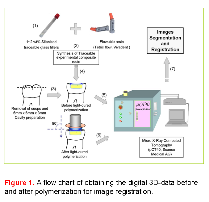
The aim of this study was to develop a method to visualize 3D polymerization shrinkage vectors analysing µCT data before and after curing light-initiated dental composites.
Materials and Methods
A high resolution micro x-ray computed tomography (µCT40, Scanco Medical AG) was used to analyse the material movement due to curing contraction of light-curing dental composites. The data were obtained with an 8 x 8 x 8 ?m³ voxel resolution. To visualize the material movement, radiolucent spherical glass fillers, average particle size of 40 ~ 70 ?m in diameter (Sigmund Linder GmbH), were chosen as traceable markers. The dimethacrylates based flowable resin (Tetric flow, Vivadent) was selected for this first experiment in order to obtain shrinkage values which can be clearly identified with the given µCT resolution. The glass beads act as traceable markers. They were silanized to ensure a durable connection to the composite. The total amount was 12 wt%. A tooth cavity, 3 mm in depth and 5 mm in diameter, was filled with the experimental resin paste. The cavities were divided into 2 groups which were treated with and without bonding agent respectively (3M, Adper Prompt L-Pop). The restoration was digitized before and after light-curing (LED SmartLight® PS, Dentsply). The 3D-data before and after polymerization were subjected to an image analysis. The 3D displacement vector field is generated with an elastic registration algorithm using an optical flow approach (Insight Segmentation and Registration Toolkit).
Results
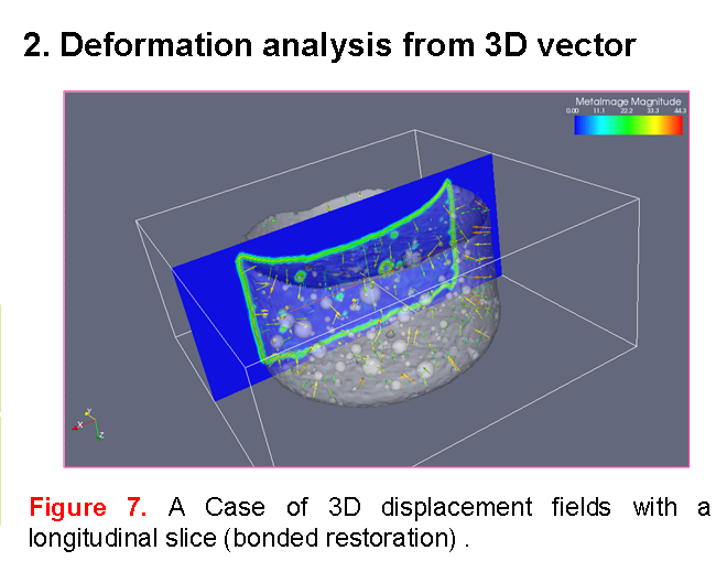
The obtained µCT images clearly showed the shrinkage vectors of light-initiated dental composites based on the material flow. The 3D displacement vector field of the µCT image illustrated that the embedded marker beads shift towards the centre of mass. The displacement field perpendicular to the z-axis confirmed this observation. In spite of bonding agent, the boundary conditions of tooth cavity affected the movement: the composite tends to adhere to one side of the cavity and a compensatory mass movement from the opposite side can be observed. For the given situations it is ascertained that the composite does not shrink towards the light source.
Conclusions
The suggested method to evaluate mass movement due to polymerization contraction can be applied to visualize the real polymerization shrinkage vectors. This method has the potential to re-evaluate all the currently available hypotheses concerning the amount and orientation of shrinkage vectors.