Visualization: Influence of endodontic therapy on the canal shape.
Root canal preparation changes the root canal anatomy. This is influenced by the operator, root canal curvature and type of instrument used. Studies to objectively quantify these changes (volume, uninstrumented root area, canal transportation) are still rare as the methods are rather complex. Therefore we developed the method ourselves. The following images represent an example how we identify and quantify the canal transportation due to endodontic therapy.
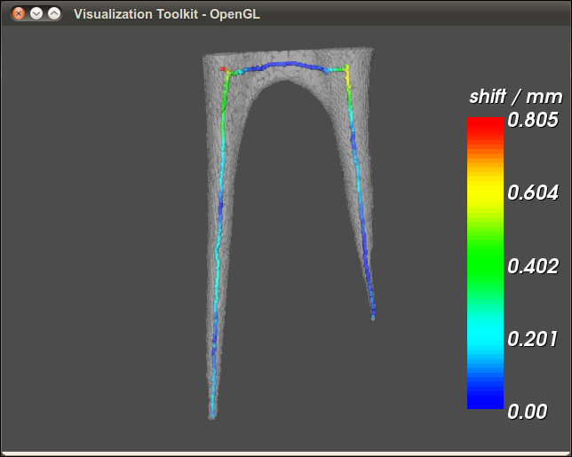
The teeth were digitized with a microCT before and after therapy. The center line of the root canal before treatment and after treatment is identified. Blue is the canal center before treatment.
After endodontic treatment. The centerline of the canal is identified again and the superimposed colors summarize the deviation from the center line before any instrumentation in mm. In the first image both canals are visible. This makes it easy to compare the results (Light blue (transparent) is the pulp cavity before treatment). The image can be turned and rotated for interactive analysis, too.
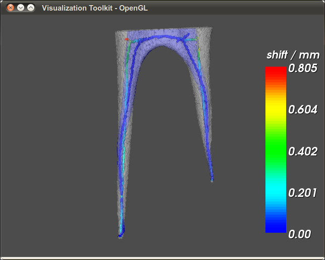
In the image below, the baseline image was removed to have a better view at the color codes. The metric data are available, too, of course.
The images are the result of a very successful cooperation between the Ludwig-Maximilians-University (Prof. Kunzelmann, Dr. D. Thiessen), the Fachhochschule Augsburg (Fakultät für Informatik, Prof. Dr. P Rösch) and the School of Dentistry, Manchester (Dr. Madarati, Prof. Dr. David Watts)